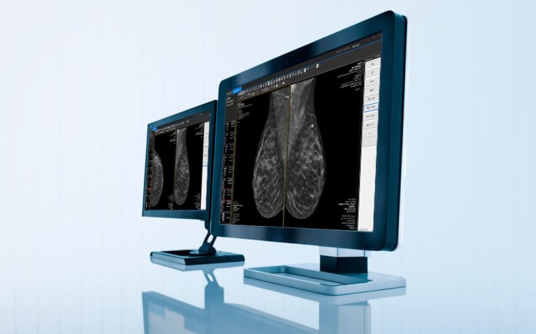
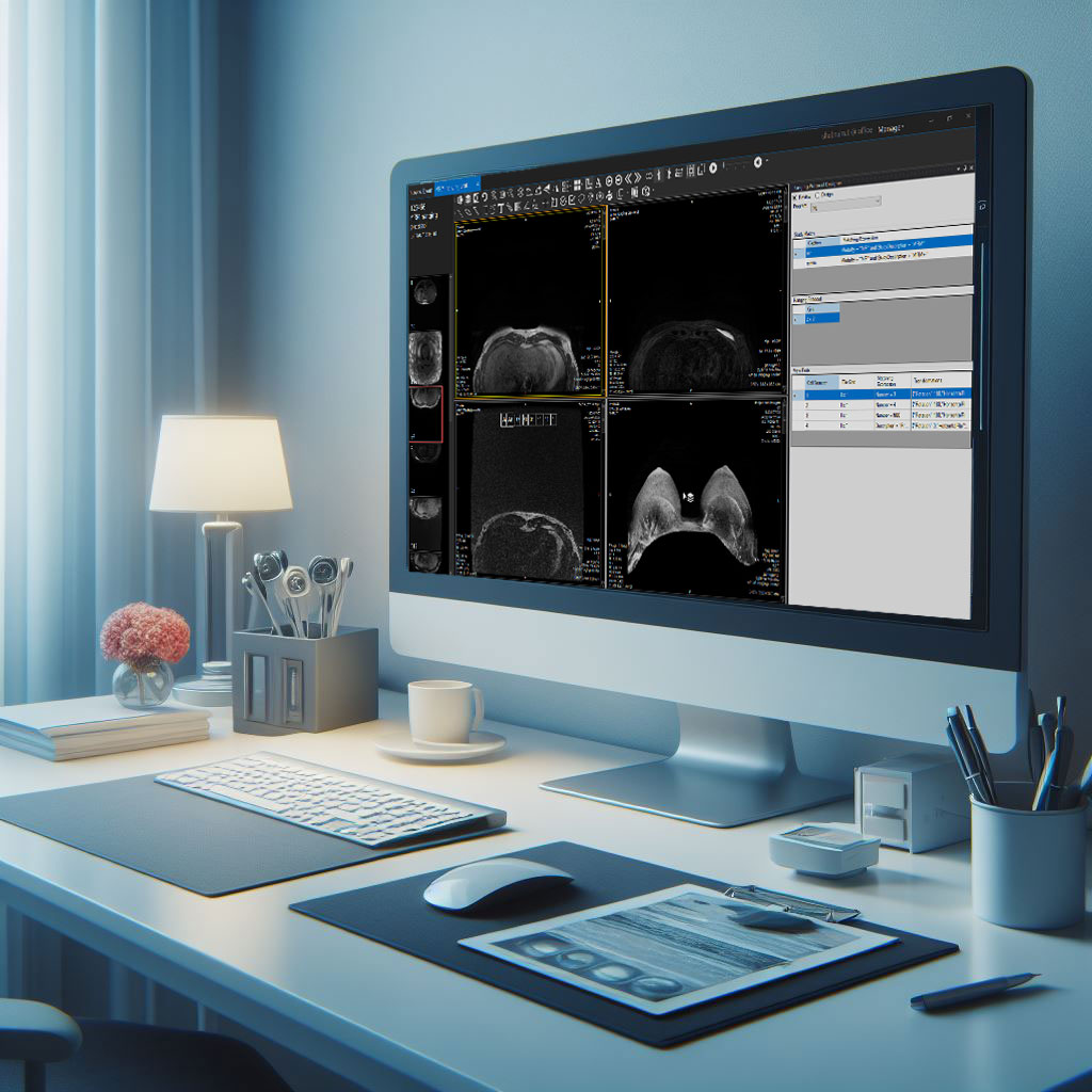
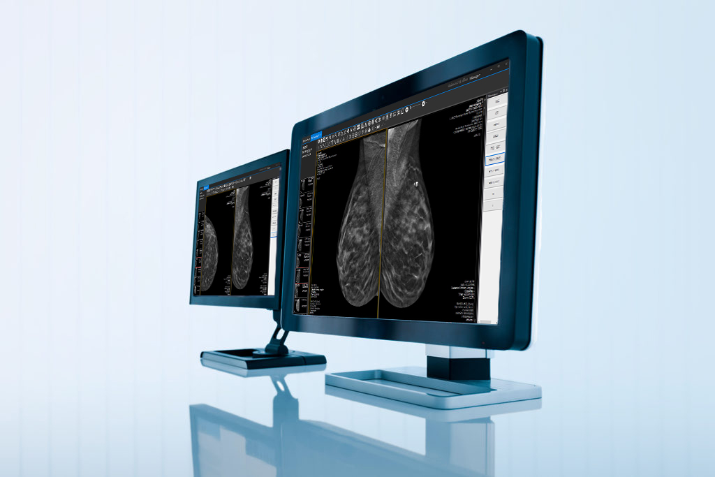
Effective mammogram hanging protocols are essential for ensuring accurate and efficient interpretation of mammographic images. Properly arranged images allow radiologists to compare current and prior mammograms, aiding in the early detection of breast cancer. This article explores the importance of mammogram hanging protocols, their key components, and the benefits they offer in breast imaging.
Why Standardized Hanging Protocols Matter
Mammogram hanging protocols ensure that images are presented consistently and organized, allowing radiologists to compare current and previous mammograms effectively. Consistent presentation is crucial because breast tissue can undergo subtle changes over time, and early detection of these changes can significantly impact patient outcomes. Standardized protocols reduce the risk of misinterpretation, minimize variability among radiologists, and improve the overall quality of breast imaging.
Key Components of Mammogram Hanging Protocols
- Image Orientation and LabelingProper orientation and labeling of mammographic images are foundational. Images are typically presented with the left breast images on the left side and the right breast images on the right side. Clear labeling includes markers for left (L) and right (R) breasts, views (craniocaudal (CC) and mediolateral oblique (MLO)), and dates of examination, ensuring radiologists can quickly and accurately identify the images.
- Comparison with Prior StudiesHanging protocols facilitate the comparison of current mammograms with prior studies, essential for detecting changes in breast tissue over time. Standardized protocols ensure that images from previous examinations are displayed alongside the current images consistently, allowing radiologists to assess changes in density, architecture, and the presence of new lesions.
- Synchronized ViewingSynchronized viewing involves presenting the same views (e.g., CC or MLO) of both breasts side by side. For example, the current CC view of the left breast is displayed next to the prior CC view of the left breast, and the same applies to the right breast. This helps radiologists directly compare corresponding images and identify differences more efficiently.
- Utilization of Digital ToolsDigital mammography has introduced advanced tools that enhance hanging protocols. Digital workstations allow radiologists to manipulate images, adjust contrast and brightness, and utilize features such as magnification and annotations. These tools aid in the detailed examination of breast tissue, ensuring radiologists can apply hanging protocols with greater precision.
Mammogram hanging protocols are indispensable tools in the field of breast imaging. By standardizing the presentation of mammographic images, these protocols enhance diagnostic accuracy, improve workflow efficiency, reduce variability, and facilitate training and education. At NESTOPIAA, we have specifically designed hanging protocols for mammogram images to ensure the highest standards of accuracy and consistency. As the field of mammography continues to evolve, the implementation and refinement of hanging protocols will remain essential for ensuring the highest standards of breast cancer detection and diagnosis. Check out Shabnam Taleghani’s insightful video on LinkedIn about mammogram hanging protocols and discover how these practices enhance breast cancer detection and diagnosis. Don’t miss it!


